[ad_1]
We knew from the beginning of the pandemic that this disease not only damages the respiratory system, it also causes chaos in the digestive system, damages the heart, causes bleeding disorders.
And now, a year after the global spread of the pandemic, careful autopsies of COVID-19 patients have shown very clearly the extent to which the virus causes inflammatory processes and damage to brain tissue. This new knowledge may help explain the abundance of neurological symptoms in some patients, from headaches, memory loss, dizziness, weakness, and hallucinations to severe seizures and strokes.
By some estimates, up to 50 percent. COVID-19 patients treated in hospitals can experience neurological symptoms that make it difficult for some people to perform everyday tasks, such as preparing food.
“We were very surprised. At first, we expected to see the damage caused by lack of oxygen. Instead, we have seen multifocal areas of injury commonly associated with strokes or inflammatory neurological diseases,” said Avindra Nathas, physician at the National Institutes of Health. (NIH) USA and Director of Clinical Activities.
NIH researchers, including physicians Myoung-Hwa Lee and A. Nath, conducted comprehensive examinations of brain tissue from 19 dead COVID-19 patients. The age of the patients ranged from 5 to 73 years, many of them were at risk due to various conditions (diabetes, heart disease).
Instead, we have seen multifocal areas of injury commonly associated with strokes or inflammatory neurological diseases.
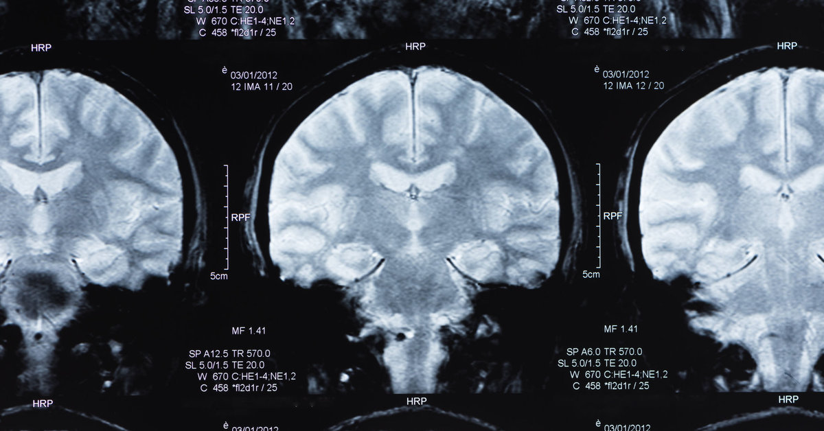
Using a powerful MRI microscope, the researchers identified 10 patients with brain abnormalities. Subsequent studies using microscopy revealed hyperintensive areas – bright areas in the micrographic image of brain samples. Fluorescence microscopy showed that these areas were rich in blood protein fibrinogen.
In many samples, the clear areas are surrounded by T cells of the immune system, as well as specialized brain immune cells, microglia. There were also dark spots: traces of clotting in the brain. As a result, the researchers concluded that these patients suffered from multiple minor brain bleeds, lesions that are characteristic of inflammatory brain diseases.
“There were very small blood vessels in the brain. And the bleeding was uneven, there were bleeds here and there ”, explained A. Nat.
The neurological symptoms of COVID-19 do not only affect the most serious patients whose lives are saved in intensive care units or patients with chronic diseases.
“We have seen a group of younger people, in addition to the usual risk factors for stroke, we have seen patients with acute mental changes and no other explanation for those changes,” said Benedict Michael, neurologist at the University of Liverpool (UK ), in September.
COVID-19 patients also experience delusions and develop psychosis. In one case, a 55-year-old woman began seeing rain and monkeys in her home and later began to think that her friends and family had been replaced by identical suitors (Capgras delusions).
Despite efforts to find traces of the virus in brain tissues, the researchers found no SARS-CoV-2 in their samples, but said in a study report:
Other studies have found virus particles in brain tissue, but these were small and the virus was rare in the brain.
“So far, our research shows that the damage we see may not be due to the fact that the SARS-CoV-2 virus directly damages the brain,” Nat said. According to him, the explanation may be our body’s inflammatory response to a viral attack.
However, due to the small number of samples used for the study and the limited amount of clinical information, the clinicians were unable to draw definitive conclusions.
Researchers are concerned that such brain damage could irreversibly change the lives of people with COVID-19: memory loss, other neurological consequences like persistent fatigue or Guillain-Barré syndrome.
The findings of the peer-reviewed study were published in the journal NEJM.
[ad_2]
