[ad_1]
A study led by the Cedars-Sinai Department of Neurosurgery has identified certain regions of the retina, the lining at the back of the eye, that are more affected by Alzheimer’s disease than other areas.
The findings can help doctors predict brain changes and cognitive decline, even for patients experiencing the first signs of mild impairment.
These clues can occur very early in the progression of Alzheimer’s disease – several decades before symptoms appear, detecting these signs can help diagnose the disease more accurately, allowing for earlier and more effective treatment intervention. .. “
Maya Koronyo-Hamaoui, PhD, corresponding study author and associate professor, neurosurgery and biomedical sciences, Cedars-Sinai Medical Center
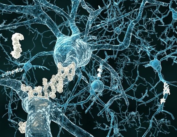
Alzheimer’s disease is the most common form of dementia, a group of brain disorders characterized by a general loss of mental abilities, including memory, judgment, language, and abstract thinking.
The findings of the new study, published in the journal Alzheimer’s and dementia: diagnosis, evaluation and monitoring of diseases, They were from a clinical trial involving people over the age of 40 who showed signs of cognitive decline.
In the trial, the researchers used a non-invasive technique known as sectoral retinal amyloid imaging to capture retinal images in the participants. The retina, which is directly connected to the brain, is the only tissue in the central nervous system accessible for non-invasive, high-resolution, patient-friendly imaging.
The images were then analyzed using a new process that could identify certain peripheral regions in the retina that best matched brain damage and cognitive status.
By studying the images, the scientists were able to detect patients with a higher accumulation of retinal amyloid protein, which means a higher probability of developing Alzheimer’s disease or cognitive impairment.
These findings are based on pioneering research in 2010 in which Koronyo-Hamaoui and her team identified a pathological hallmark of Alzheimer’s disease, deposits of beta amyloid protein, in retinal tissues of deceased patients.
The team then developed a methodology to detect amyloid beta protein plaques in the retina in living patients with the disease.
In another research study involving laboratory mice, which was recently published in the journal Aging Cell, Koronyo-Hamaoui, premedication student Jonah Doustar and other members of the research team further validated the role of the retina. in the visualization of distinctive signs of Alzheimer’s disease. and identified a potential treatment to combat the disease.
“We found that increased levels of beta-amyloid peptides in the retina correlated with levels found in brain tissues, even in the later stages of Alzheimer’s disease,” Koronyo-Hamaoui said.
“We also suggest a particular type of immune modulation therapy that can fight disease by reducing toxic proteins and damaging inflammation in the brain and, in return, enhancing a type of protective immune response that preserves the connections between neurons, which are closely connected to cognition. “
Both studies show promise for diagnosing and treating Alzheimer’s disease, a condition that affects more than 5.5 million people in the U.S., said Keith Black, MD, professor and chair of the Department of Neurosurgery.
“This work may guide future brain and retina imaging studies to detect Alzheimer’s disease, assess disease progression, and identify early treatment options,” Black said.
Source:
Cedars-Sinai Medical Center
Magazine reference:
Dumitrascu, OM, et al. (2020) Sector segmentation of retinal amyloid images in subjects with cognitive impairment. Alzheimer’s and dementia: diagnosis, evaluation and monitoring of diseases. doi.org/10.1002/dad2.12109.
