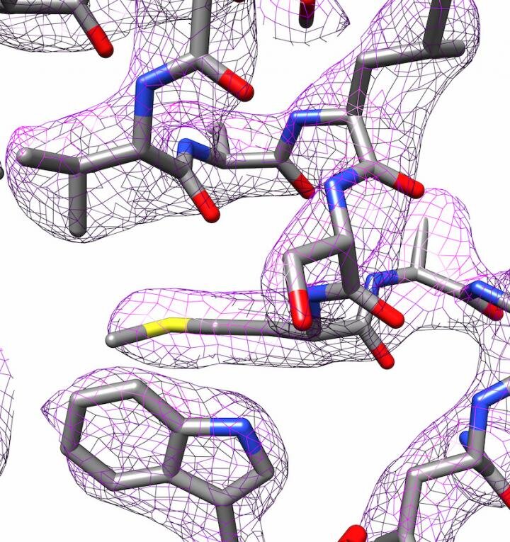

3D structure of apoferritin produced by electron cryoscopy. Credit: Panagiotis Kastritis
Biochemists at Martin Luther University in Halle-Wittenberg (MLU) have used a standard electron microscope to produce surprisingly good images that are on par with those taken by much more sophisticated equipment. They have managed to determine the structure of ferritin almost at the atomic level. Their results were published in the magazine. MORE ONE.
Electronic cryoscopy has become increasingly important in recent years, especially to shed light on protein structures. The developers of the new technology received the Nobel Prize in Chemistry in 2017. The trick: samples freeze instantly and then bombarded with electrons. In traditional electron microscopy, all the water is first drawn from the sample. This is necessary because the research is carried out in a vacuum, which means that the water will evaporate immediately and make imaging impossible.
However, because water molecules play such an important role in biomolecules, especially proteins, they cannot be examined with traditional electron microscopy. Proteins are among the most important building blocks of cells and perform a variety of tasks. A deep understanding of their structure is necessary to understand how they work.
The research group led by Dr. Panagiotis Kastritis, group leader at the HALOmem Center for Innovation Competency and junior professor at the Institute of Biochemistry and Biotechnology at MLU, purchased a state-of-the-art cryoscopy microscope in 2019. “No there’s another microscope like this in Halle, “says Kastritis. The new Thermo Fisher Glacios 200 kV, funded by the Federal Ministry of Education and Research, is not the best and most expensive microscope of this type.
However, Kastritis and colleagues were able to determine the structure of the iron storage protein apoferritin up to 2.7 ångströms (Å), in other words, almost down to the individual atom. An ångström equals one tenth of a nanometer. This places the research group in a league similar to departments with much more expensive equipment. Apoferritin is often used as a reference protein to determine the performance levels of the corresponding microscopes.
Recently, two research groups broke a new record with a resolution of approximately 1.2 Å. “Such values can only be achieved using very powerful instruments, which only a handful of research groups around the world have at their disposal. Our method is designed for microscopes found in many laboratories,” explains Kastritis.
Electronic cryoscopes are very complex devices. “Even small misalignments can make images useless,” says Kastritis. It is important to program them correctly and Halle has the technical experience to do so. But the analysis that is done after the data has been collected is just as important. “The microscope produces several thousand images,” explains Kastritis.
Image processing programs are used to create a three-dimensional structure of the molecule. In cooperation with Professor Milton T. Stubbs of the MLU Institute of Biochemistry and Biotechnology, the researchers have developed a new method for creating a high-resolution protein model. Stubbs’ research group uses X-ray crystallography, another technique for determining protein structure, that requires proteins to crystallize. They were able to combine a modified form of an image analysis technique with images taken with the electron microscope. This made the charge states and the individual water molecules visible.
“It is an attractive method,” says Kastritis. Instead of needing very expensive microscopes, it requires a lot of computing power, which MLU has. Now, in addition to using X-ray crystallography, electron cryoscopy can be used to produce images of proteins, especially those that are difficult to crystallize. This allows collaboration, both inside and outside the university, in the structural analysis of samples with medical and biotechnological potential.
Realization of a new image-based structure analysis method for three-dimensional structural analysis of biology.
Farzad Hamdi et al, 2.7 Å M-musculus H-chain apoferritin cryo-MS structure vitrified from a compact 200 keV microscope cryoscope, MORE ONE (2020). DOI: 10.1371 / journal.pone.0232540
Provided by Martin-Luther-Universität Halle-Wittenberg
Citation: Cryo-electron microscopy: use of inexpensive technology to produce high-resolution images (2020, July 13) retrieved on July 14, 2020 from https://phys.org/news/2020-07-electron-cryo-microscopy -inexpensive-technology- high-resolution.html
This document is subject to copyright. Other than fair dealing for private study or research purposes, no part may be reproduced without written permission. The content is provided for informational purposes only.
