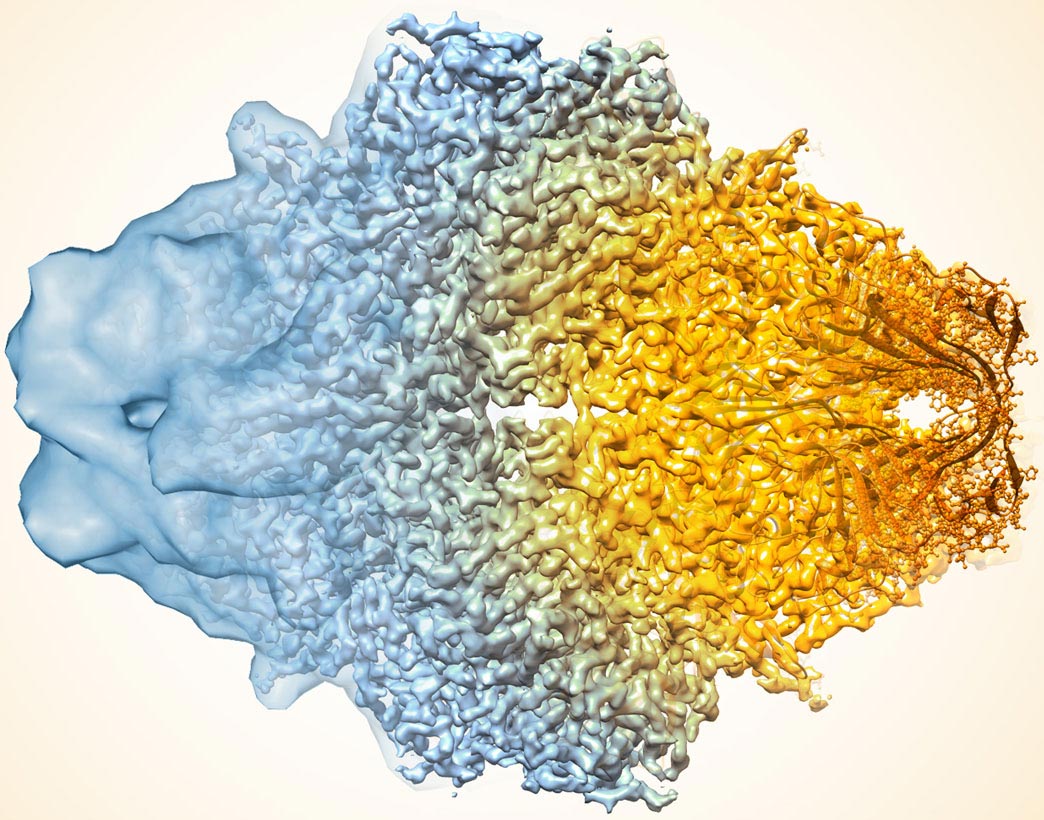

A composite image of the enzyme lactase, showing that the resolution of cryo-EM has improved dramatically in recent years. Older images on the left, latest on the right. Credit: Veronica Falconieri / National Cancer Institute
Scientists have developed a technique that improves the resolution of cryo electron microscopy.
We saw that moment on a Cop TV show where a detective is reviewing granular, low-resolution security footage, spotting someone with an interest on the tape and telling CSI technology to “enhance it”. After a few keyboard clicks, and voila – they’ve got a complete, clear picture of the suspect’s face. This, of course, does not work in the real world, as many film critics and people on the Internet prefer to point out.
However, real-life scientists have only recently developed a “growth” tool: one that improves resolution and Accuracy Powerful microscope used to demonstrate biological medicine and medical insights.
In a study published in Methods of nature, Led by Tom Terviliger of the New Mexico Consortium and a multi-institutional team, including researchers from Lawrence Berkeley National Laboratory (Berkeley Lab), showcases the new computer algorithm in cryo-electron microscopy (Cryo) generated by a 3D molecule. -EM).
For decades, these cryo-EM maps – generated by taking many microscopic images and using image-processing software – have been crucial tools for researchers to learn how the molecules of animals, plants, microbes and viruses work. And in recent years, cryo-EM technology has advanced to the stage where it can produce structures with atom-level resolution for many types of atoms. Yet in some situations, even the most sophisticated cryo-EM methods produce maps with lower resolution and more uncertainty than the need to strip the details of complex chemical reactions.

Using the enzyme β-galactosidase, also called lactase, as a test case, the researchers applied standard methods (a) and then (b) without correction algorithms and with filters to improve the uniformity of sound in the map), both The map is more similar to the submitted high-resolution protein structure map (D). Credit: Terviliger et al. / Nature methods
“In biology, we gain a lot by knowing the structure of molecules,” said study co-author Paul Adams, director of the Department of Molecular Biophysics and Integrated Bioimaging at the Berkeley Lab. “The improvements we see with this algorithm will make it easier for researchers to determine atomistic structural models from electron cryo-microscopy data. This is especially the result for modeling very important biological molecules, such as those involved in the transcription and translation of genetic code, which mostly appear only in low-resolution maps due to their complex and multi-unit structures. “
The algorithm sharpens the molecular map by filtering the data based on current knowledge of what the atom looks like and how to best estimate and remove noise (unwanted and irrelevant data) in microscopy data. An approach with the same theoretical basis was previously used to improve the structure of structures derived from X-ray crystallography, and scientists have previously suggested its use in cryo-EM. But, according to Adams, no one has so far been able to show specific evidence that Cryo-EM works.
A team of scientists from the New Mexico Consortium, Los Alamos National Laboratory, Baylor College of Medicine, Cambridge University and Berkeley Labs first used the algorithm on a publicly available map of the human protein Apoferitin, which is believed to be 1.1-enzyme. Resolution (Angstrom is 10 billionths of a meter; for reference, diameter of carbon) Atom 2 is estimated to be angostrom). They then compared their enhanced version with another publicly available apopheritin reference map with a 1.8-angstrom resolution and found an improved correlation between the two.
Next, the team used their approach on 104 map datasets of an electron microscopy data bank. For a large proportion of this map set, the algorithm improved the relationship between the experimental map and the known atomic structure and increased the visibility of the details.
The authors note that the obvious advantage of the algorithm in disclosing important details in the data, its ease of use – it is an automated analysis that can be performed on a laptop processor – will probably make it part of its standard move beyond the Cryo-EM workflow. In fact, Adams has already added the source code of the algorithm to the Phoenix software software suite, a popular package for automated macromolecular structure solution, for which he led the development team.
The research was part of Berkeley Lab’s pioneering efforts to advance the capabilities of cryo-EM technology and its use for basic science discovery. Scientists at the Berkeley Lab have been involved in a number of progressive discoveries that have enabled the development of Cryo-EM and later led it to its extraordinary current resolution.
Reference: Thomas C. Terviliger, Steven J. Ludtek, Randy J. Reed, Paul de ams dums and Powell v. “Improving Cryo-EM Maps by Changing Density” by Ephone Methods of nature.
DOI: 10.1038 / s41592-020-0914-9
