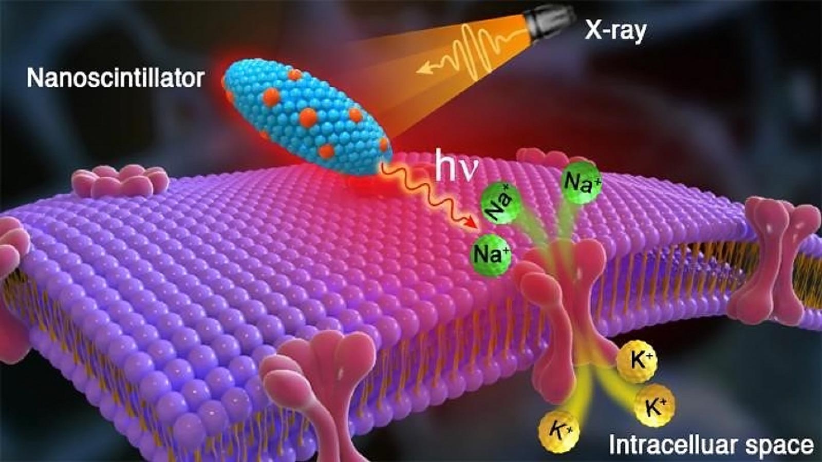

The artist’s rendering shows X-ray striking radioluminescent nanoparticles in the brain, emitting red light that stimulates the flow of sodium (Na +) and potassium (K +) ions and activates brain neurons there. Credit: Image by Zhaoi Chen / Argone National Laboratory.
Scientists are discovering a method for wireless modulation of neurons with X-rays that could improve the lives of patients with brain disorders. The X-ray source is just as necessary as the machine found in the dentist’s office fee.
Many people around the world suffer from movement-related brain disorders. The incidence of epilepsy is high 50 Million; Essential vibration, 40 Million; And Parkinson’s disease, 10 Million.
Relief for some brain disorder sufferers Could come in the form of a new treatment discovered by researchers from the Department of Energy (Please) Argonne National Laboratory and four universities. Treatment is based on both optics and the development of heredity. It will apply not only to movement-related brain disorders, but also to severe depression and pain.
“Our precision high-precision nonviolent approach can be regularized using small X-ray machines, which are commonly found in dental office fees.” – Elena Rozkova, nano scientist at Argo’s Center for Nanoscale Materials
This new treatment involves stimulation by injected nanoparticles of neurons inside the brain that are released when in contact with X-rays (nanosyntilators) and will eliminate the invasive brain surgery currently in use.
“Using a small X-ray machine can make it common for us to adopt a high-precision route, which is common in dental office fees. ”C.N.M.), a Please Office of Science User Facility.
Disorders of conventional deep brain stimulation require an invasive neurosurgical procedure when conventional drug therapy is not an option. U.S. In a traditional procedure approved by the Food and Drug Administration, surgeons place a calibrated pulse generator under the skin (similar to a pacemaker). It then connects to the insulated extension cord with electrodes inserted into a specific area of the brain to stimulate the surrounding neurons and control abnormal impulses.
“Spanish-American scientist Jose Manuel Rodriguez Delgado famously demonstrates deep brain stimulation in bullying 1960O, ”said Vasily Tytsarev, a neurobiologist at the University of Maryland and co-author of the study. And“Sending a radio signal to the implanted electrode, it stopped charging a raging bull. “
About 15 Years ago, scientists introduced the revolutionary neuromodulation technique,“to pathogenetics, ”which depends on the genetic modification of certain neurons in the brain. These neurons form a light-sensitive ion channel in the brain and, from there, ignite in response to external laser light. This approach, however, requires very thin fibroptic wires implanted in the brain and suffers from the limited depth of penetration of laser light through biological tissues.
The team’s alternative to pathogenetics approach uses a nanosynthetizer injected into the brain by bypassing implantable electrodes or fibroptic wires. Instead of lasers, they substitute X-rays, due to their greater ability to pass through biological tissue barriers.
“Injected nanoparticles absorb X-ray energy and convert it into red light, which has a significantly greater depth than blue light, “said Zhao Chen, the former. C.N.M. Postdoctoral fellow.
“Thus, nanoparticles serve as an internal light source that makes our system work without wires or electrodes, ”Rozkova added. The team’s approach can stimulate both targeting and stimulating small areas, so Rozkova noted that it has applications other than brain disorders. For example, it can be applied to heart problems and other damaged muscles.
One of the keys to success was the collaboration between two world-class facilities at Argo: C.N.M. And argon’s advanced photon source (APS), a Please Office of Science User Facility. Work on these features began with the synthesis and multi-tool characterization of nanosyntilators. In particular, X-ray excited optical luminescence of nanoparticle samples APS Beamline (20-B.M.). The results showed that the particles were extremely stable during months and upon repeated exposure to high-intensity X-rays.
According to Zoo Finfrock, the staff scientist APS 20-B.M. Beamline and Canadian light source,“They shone a beautiful orange-red light. ”
Next, Argo sent a CNM-manufactured nanosyntilator to the University of Maryland for rat tests. A team from the University of Maryland performed the tests in two months with a small portable X-ray machine. The results proved that the process worked as planned. To respond to the red light to respond to X-ray pulses with brain waves recorded on an electroencephalogram, whose brains were genetically modified.
Eventually, a team from the University of Maryland sent Ergo to the animal’s brain for characteristics using X-ray fluorescence microscopy performed by scientists. The analysis by Olga Antipova Microprobe Beamline (2-ID-E) On APS And hard X-ray nanoprobes by Zongho K (26-ID) Jointly operated C.N.M. And APS.
This multi-instrument configuration made it possible to see tiny particles living in a complex environment of brain tissue with a super-resolution of dozens of nanometers. It also allowed visualization of near and far neurons from the injection site on a microscale. The results proved that the nanocontroller is chemically and biologically stable. They do not deviate or degrade from the injection site.
“Sample preparation is very important in this type of biological analysis, ”said Antipova, a physicist in the X-ray Division.XSD) At APS. Antiopova was assisted by Kiaoling Jin and Xueli Liu, who designed some parts of the thick brain like a jeweler. Accuracy.
“There is a strong level of professional interest in pathogenetics for medical applications, ”Rozkova said. And“Although still in the proof-of-concept stage, we predict that our patent-pending wireless approach to small X-ray machines should have a bright future. “
Reference: “Wireless to pathogenetic modulation of cortical neurons enabled by radioluminescent nanoparticles.” Antipova, Zhongho K, Hiroyoki Arkawa, Fritz W. Hrichikan, Rosen Da Dong Ngi Wang, Yi Liu, Brandon Gatin, Yang Tao, Yu Chen, Reha S. Erzurmalu, Huango Yang and Elena A. Rozkova, 24 February 2021, ACS Nano.
DOI: 10.1021 / acsnano.0c10436
Related Articles“Wireless to pathogenetic modulation of cortical neurons enabled by radioluminescent nanoparticles ” ACS Nano. In addition to Rozkova, Chen, Finfrock, Antipova and Kai, other Argos are author Rosemary Wilton. University contributors include Vasily Taisterev, Da Dong Ngi Wang, Yi Liu, Brandon Gaitan, Yang Tao, and Yu Che, Department of Bioengineering, University of Maryland; Hiroyuki Arkawa and Reha Erzurumulu from the University of Maryland School of Medicine; Fritz Lischa from Uniform Services University of Health Sciences; Brian Hooks, Department of Neurobiology, University of Pittsburgh; And Huango Yang from Fuzhou University.
This research was supported Please Office of Science, National Institutes of Health and National Science Foundation.
