A mysterious network of white blood cells that can attack and destroy cells infected with the coronavirus has taken on new importance as epidemiologists continue their search for a vaccine in doubt that antibodies can only provide lasting immunity.
These blood cells, called T cells, act as snipers in a plateau of soldiers of the immune system as they stalk and kill infection. It is a microscopic skill set that scientists hope to use against SARS-CoV-2, the specific coronavirus that causes COVID-19.
The human body, in fact, is waging a constant war against invaders, and the production of a vaccine against the coronavirus will require molecular biologists to utilize these weapons. This includes B cells, which secrete antibodies that neutralize pathogens, and T cells, which destroy the pathogens and any toxic molecules they produce.
T cells have been thrown into the limelight, as recent studies suggest that the human body may not be able to retain antibodies produced by B cells for very long, raising questions about whether permanent immunity to COVID-19 is possible after people recover them.
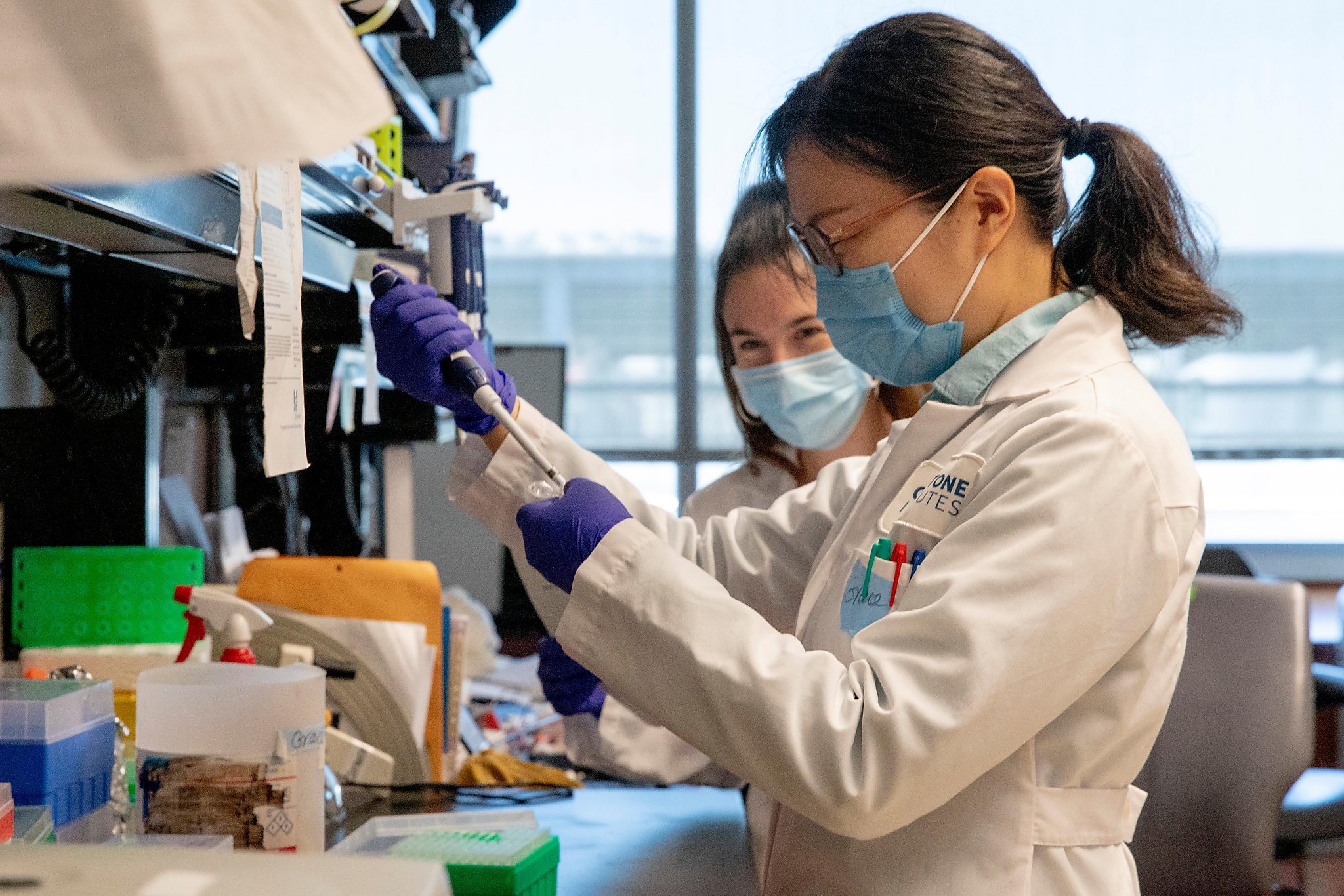
Nadia Roan, an associate professor at UCSF and a T-cell expert, said the key to a vaccine could be to figure out how to inspire T-cells to target the coronavirus even in the absence of antibodies, the workhorses of the human immune system.

Roan and her colleagues found that a robust population of T cells attacking the coronavirus progresses to mild infections, and these cells persist for months and can multiply. The results, which were accepted for publication on August 14 in the peer-reviewed journal Cell Reports Medicine, suggest that T cells may play an important role in the fight against the virus.
“We found a diverse collection of T cells that recognize SARS-CoV-2, each serving its own specific functions,” Roan said. “Importantly, these T cells existed more than two months after recovery from infection, and were able to increase markedly in number.”
The UCSF’s findings were similar to those found by researchers at France’s Strasbourg University Hospital in a preliminary study recently published in medRxiv, an online service distributing unpublished manuscripts. Both studies found that the T response was observed for at least 69 days in patients recovering from mild cases of COVID-19.
A Swedish study published last week in the scientific journal Cell found SARS-CoV-2-specific T cells in patients, including asymptomatic individuals, who had no detective antibodies.
The Roan research team plans to monitor T-cell responses in patients for six months to see if they are still present.
“It’s kind of encouraging,” said Roan, who works in the UCSF urology department and is a researcher for the Gladstone Institutes, an independent biomedical research organization.
The studies are an example of the complexities that scientists face in their accelerated search for a vaccine for coronavirus.
Of the 150 potential vaccines for coronavirus at various stages of development, at least five elicited anti-anti and T-cell responses. These include RNA vaccines developed by Massachusetts biotechnology company Moderna and New York City firm Pfizer.
The ability to stimulate T cell production, as well as replication, is expected to be a crucial part of any vaccine being developed. Roan and her colleagues recently published a separate study showing that an egg white called Interleukin-7 stimulated the number of T cells. Interleukin-7 treatment is also undergoing a clinical trial in the United Kingdom.
“It is good news for us that these cells that can fight SARS-CoV-2 can expand in numbers,” she said.
Researchers must also find out how T can stimulate cell growth without overreacting to the immune system. T cells, when stimulated too much, can attack healthy cells. Doctors say that overreactive immune responses, called cytokine storms, have been responsible for many of the most serious cases of COVID-19 and for quite a few deaths.
Inserting T cells is not easy, given that researchers do not understand all the ways of the virus with the human body. The orbit requires a deep dive into the diverse collection of cells, amino acids and proteins that make up the human immune system.
There are two types of T cells – known as CD4 and CD8. CD4 cells are known as helpers because they help all other cells, including the production of antibodies by B cells. The CD8 cells are killers of the body, lying dormant in the bone marrow and lymphoid tissues, where they jump out and attack infected cells, according to microbiologists.

It is possible to distinguish between the two using technology called massacytometry, Roan said. In the past, scientists collected blood cells and analyzed them all mixed like a fruit smoothie. Mass cytometry allows each individual T cell to be identified, measured and marked with some sort of ion barcode.
Roan and her colleagues used the technology to track the slow increase in CD4 and CD8 T cells in a single male COVID-19 patient with lymphopenia, extremely low white blood cell counts common in severely ill coronavirus patients. They observed a large increase in CD4 helper cells after 26 days and only a hint of CD8 attackers. Then, 14 days later, both types of T cells were found in abundance.
The complete T-response, although delayed, was so effective that the man was released from the hospital.
“It suggests that T cells may play a more beneficial role in helping with recovery,” Roan said.
But T cells are only part of the complex picture. The invasive virus binds to the ACE2 receptors of healthy cells, breaks in and replicates. The body’s immune response flags infected cells by placing fragments of protein, such as peptides, on their surface, according to experts. These peptides inspire T cells to break down into action, which in turn can help B cells make antibodies. Then antibodies can attach to the virus and prevent it from binding to and entering other healthy cells.
Antibodies are also designed by B-cells to match an invading virus in a very specific way, allowing them to bind tightly and neutralize viral functions. Typically, the stronger the binding, the more effective the antibody is in preventing the pathogen from entering the cell, according to infectious disease specialists.
However, viruses are able to mutate in ways that can remove this perfect match, thereby reducing the effectiveness of antibodies. The flu virus often mutates, which is why people need a different flu every year.
And antibody levels can decrease over time, leaving too few of them to block all virus particles. That may be another reason why people need booster photos with some infectious diseases.
When human antibodies lose their effectiveness in these ways, it can also impair the ability of scientists to produce effective vaccines. This is because vaccines are designed to produce viral proteins, called antigens, which in turn stimulate the human immune system to respond, including through the production of antibodies.
Vaccines can also call for the help of T cells, which recognize infected cells that do not protect the antibodies.
“T cells can provide protection completely absent from antibodies. They help provide a stronger response, ”said Jason Cyster, a professor in the Department of Microbiology and Immunology at UCSF. “T cells specific for the virus proliferate once they are exposed to the virus and then become aggressive and attack the infected cell.”
One goal of many fax developers is to figure out how to take advantage of these unique capabilities. The key to this can be found in the ability of human T cells to develop a kind of memory.

After most T cells are done fighting an infection, a small population of ‘memory’ T cells is left behind, according to Roan. The numbers of these cells, and how long they last, can vary, but these cells have the powerful ability to reappear when the same virus strikes again.
“If in the future the person is infected with the same virus, these T memory cells can respond faster and more effectively than the first time,” Roan said. “That’s in part how faxes work, by generating memory cells.”
The phenomenon can also occur with B cells.
Dr Jay Levy, a specialist in immunology and virology and a professor of medicine at UCSF, said that cerebral palsy is the reason why the smallpox vaccine has been working for decades, even though the antibody response is inoculated 75% after six months to humans. .
A recent paper found that patients who contracted SARS, a coronavirus identified in China in 2003, had long-lasting T-cell immunity for at least 17 years after being infected, Levy said.
Immunity decreases with some diseases, he said, and that is why booster shots are sometimes needed for, among other things, polio. Repeated exposure to certain childhood infections, such as chickenpox and mumps, naturally stimulates immunity, he said.
“The original B cell that produces the antibodies remains round, probably in the bone marrow, but probably in other places, such as the lymph nodes, as B cells for memory,” Levy said. “If the antibody starts to decrease, it does not mean that the person cannot react when the agent comes in again.”
Scientists are focusing on maximizing the effectiveness of these memory cells as they try to develop vaccines. But Roan said SARS-CoV-2 is uncommon and, for some individuals, the memory response does not appear as predicted.
“We want to elicit a comprehensive immune response,” Roan said. But “as the antibody response grows, the T cells may play an important role.”
Peter Fimrite is a staff writer for the San Francisco Chronicle. Email: [email protected] Twitter: @pfimrite