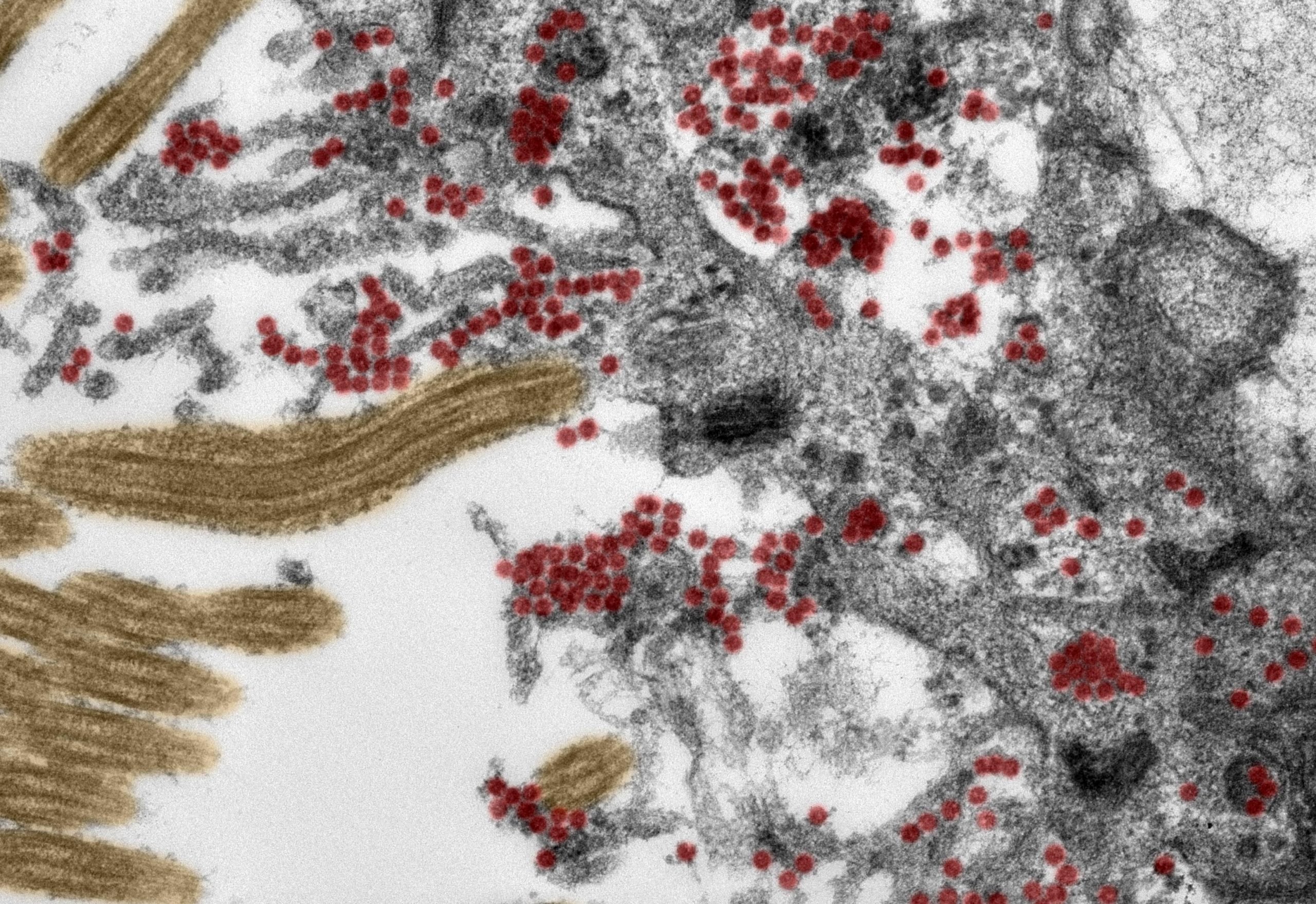

An electron microscope image (ultrathin section, artificially colored) shows a section of decorated cells in the olfactory mucosa. Intact SARS-CoV-2 particles (red) are found in large numbers both inside the cell and on cellular processes. Yellow: Kinosilia. Credit: Michael Law / RKI and Carsten Dittmeyer / Charita
Using post-mortem tissue samples, a team of researchers Charita – Universitismetizin Berlin Has studied the mechanisms by which the novel coronavirus can reach patients’ brains COVID-19, And once the immune system reacts to the virus, it is gone. The results, which show that SARS-CoV-2 The olfactory mucosa enters the brain through nerve cells, have been released Nature Neuroscience. For the first time, researchers have been able to produce electron microscope images of intact coronavirus particles inside the olfactory mucosa.
It is now recognized COVID-19 It is not a completely respiratory disease. In addition to affecting the lungs, SARS-CoV-2 Can affect the cardiovascular system, gastrointestinal tract and central nervous system. With more than one in three people COVID-19 Report neurological features such as loss of their smell or taste, headache, fatigue, dizziness and nausea. In some patients, the disease can lead to stroke or other serious conditions. Until now, researchers have suspected that these manifestations may be due to viruses entering and infecting specific cells in the brain. But how does SARS-CoV-2 Go there Under the joint leadership of Helena Ruddbrush CharitaProf. of the Department of Neuropathology and the Director of the Department. Dr. Frank. Frank Hepner, a multidisciplinary team of researchers, has now discovered how the virus enters the central nervous system and then invades the brain.

Immunofluorescence staining shows nerve cells (pink) inside the olfactory mucosa that have been infected with SARS-Cavi-2 (yellow). The epithelial cells appear blue. Credit: Jonas Franz / Universitismetzin G ગttingen
As part of this research, experts in the fields of neuropathology, pathology, forensic medicine, virology and clinical care studied tissue samples from 33 patients (average age 72૨) who either died. Charita Or University Medical Center Göttingen after signing the contract COVID-19. Using the latest technology, the researchers analyzed samples taken from the olfactory mucosa of dead patients and from four different areas of the brain. Both were tested for tissue samples and specialized cells SARS-CoV-2 Genetic material and ‘spike proteins’ that are found on the surface of the virus. The team provided evidence of the virus in various neuroanatomical structures that connect the eyes, mouth, and nose to the brain stem. The olfactory mucosa revealed the highest viral load. Using special tissue stains, the researchers were able to produce first-class electron microscopy images of intact coronavirus particles inside the olfactory mucosa. Both of these were found both in nerve cells and in extended processes from nearby accessory (epithelial) cells. All samples used in this type of image-based analysis should be of the highest quality. To confirm this case, the researchers ensured that all clinical and pathological procedures were closely coordinated and supported by a systematic infrastructure.
“These data support the notion that SARS-CoV-2 The brain is able to use the olfactory mucosa as an entry port, says Professor Hepner. This area is also supported by the close formation of mucosal cells, blood vessels and nerve cells. The neuropathologist adds, “Once in the olfactory mucosa, the virus uses neuroanatomical connections, such as the olfactory nerve, to reach the brain. “Nevertheless, it is important to emphasize that COVID-19 What was defined as a serious disease in the patients involved in this study is associated with a small group of patients in whom the disease proves to be fatal. Therefore, it is not possible to translate the results of our study into mild or moderate disease cases. “
The way the virus moves from the nerve cells is fully explained. “Our data suggest that the virus travels from nerve cells to nerve cells to reach the brain,” explains Dr. Redburch. He goes on to say: “However, the virus is also carried by blood vessels, as evidence of the virus has also been found in the walls of blood vessels in the brain.” SARS-CoV-2 Is far from the only virus capable of reaching the brain through certain routes. “Other examples include the herpes simplex virus and the rabies virus,” explains Dr. Redburch.
The researchers studied how the immune system responds to infection SARS-CoV-2. In addition to finding evidence of active immune cells in the brain and olfactory mucosa, they found immune signatures of these cells in the brain fluid. In some of the cases studied, researchers have also found tissue damage due to stroke as a result of thromboembolism (i.e. blockage of blood vessels due to blood clotting). “In our eyes, the presence of SARS-CoV-2 Provides good descriptions of olfactory mucosa nerve cells, similar to neurologic symptoms COVID-19 Patients, such as loss of sense of smell or taste, “explains Pro Hepner.” We also found SARS-CoV-2 In those areas of the brain that control important functions such as respiration. It cannot be ruled out, in severe patients COVID-19, The presence of the virus in these areas of the brain will have a more severe effect on respiratory function, due to shortness of breath SARS-CoV-2 Lung infections. Such problems may also arise in relation to cardiovascular function. “
References: Jenny Meinhart, Josephine Redke, Carsten Dittmeyer, Jonas Franz, Carolina Thomas, Ronza Mothes, Michael Lau, Julia Snyder as “Office Factory Transmucosal SARS-CoV-2 Invasion of Individuals with COVID-19”. , Sebastian Brink, Selena Greel, Malte Lehmann, Olga Hassan, Tom Ashman, Alyssa Schumann, Robert Lorenz Chua, Christian Conrad, Roland Isles, Werner Stanzel, Mark Windgassen, Larissa Rößller, Hans-Hell Andres Nietzsche, Walter J. Schulz-Schaefer, Sami Hakrosh, Martin S. Winkler, Byrne Tempe, Franziska Skeeb, Peter Cartwilice, Dirk Reinhold, Britta Sigmund, Anja a. Cahill, Sefer Elizkerts, Devil Horse, Barbara Ingold-Hepner, Christine Stadelman, Christian Droston, Victor Max Corman, Helena Rudbrich and Frank L. Hepner, 30 November 2020, Nature Neuroscience.
DOI: 10.1038 / s41593-020-00758-5
