[ad_1]

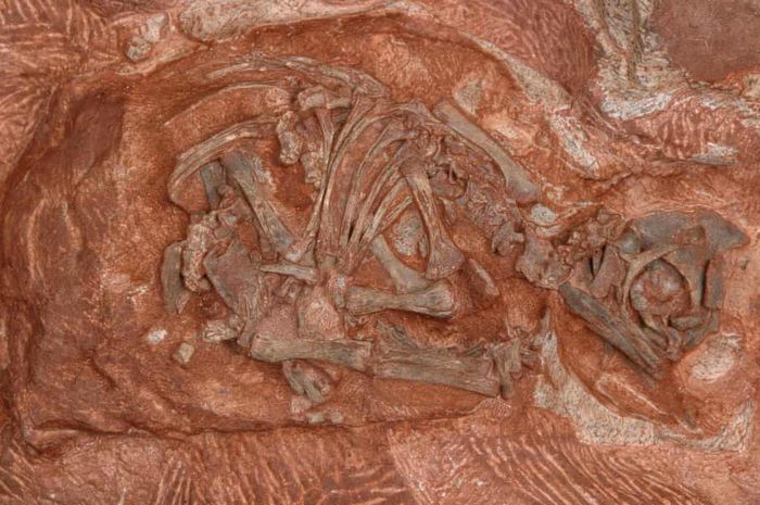
Skeletal dinosaur embryo Massospondylus.
Nationalgeographic.co.id – The dinosaur fossil skulls that died in their eggs some 200 million years ago have been digitally reconstructed by scientists. This sheds new light on the development of the animals and how close they are to hatching.
Egg fossils called The rare clutch of seven eggs …some contain embryos, discovered in South Africa in 1976, along with young creatures that are developing as a species of dinosaur called Massospondylus Carinatus.
Plant eaters are the ancestors of sauropod dinosaurs like diplodocus and will walk on two legs when they grow up. They are about five meters from nose to tail, and have a long neck with a small head.
Also read: ‘Broken Hill’ Skull assesses the process of modern human evolution
Now investigators say they have Computed tomography High resolution to digitally reconstruct a small skull from an embryo.
Previous attempts to discover how adult dinosaur embryos, such as observing the level of contact between various parts of the skull, had difficulties, especially due to the challenge of evaluating these characteristics. However, according to Dr. Kimi Chapelle, a researcher at the University of the Witwatersrand who participated in the study, a scan It offers a different approach, allowing researchers to see the extent of bone formation and explore parts of the sample that are hidden from view.
New research published in the magazine. Scientific reports, revealing the remaining embryos in three eggs, as well as the skull showing two sets of teeth.
Even so, While one set shows teeth and is similar to adult teeth, as seen previously in other dinosaur embryos, the other sets are different, consisting of simple cone-shaped teeth. “We have never seen that before,” Chapelle said, adding that, like many reptiles today, dinosaurs could lose or reabsorb these teeth before developing teeth that would hatch.
The team also took previously collected data on embryos from three live animals today: African turtles, chickens, and Nile crocodiles, and tracked the location and rate of bone tissue formation in the embryo during incubation. They then applied their findings to another living animal, the central bearded dragon.
The results showed that although the animals had different incubation times, the relative sequence and time of bone tissue formation with the skull were similar.
Featured videos
PROMOTIONAL CONTENT
[ad_2]