If you have ever studied any chemistry or biological studies, there is a very good chance that you will be able to present a general picture of what is thought to be a chromosome.
Millions of high-schoolers and undergraduates will be convinced that it is a tall, narrow X-shape – imagining what the two joined chromatids look like after DNA replication, but before cell division is complete, at what stage did they separate? Have their own individual chromosomes.
Unfortunately, this is a minor problem with ubiquitous symbols, scientists say, at least in terms of how accurate their representation is.
“For 90 percent of the time, chromosomes don’t exist,” says Jun-Han Sue, a former physician-scientist at Harvard University. “
In a study published this year, Sue and her team devised a new way of imaging the 3D organization of chromatin in human cells, giving us a better understanding of chromosome chemistry than even the iconic X.
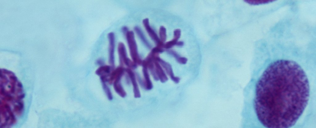
 (Xiaoi Zhuang Lab)
Above: Multicolor image of crotmatin, in situ hybrid and using multiplexed fluorescence in super-resolution microscopy.
To understand the molecular mechanisms within an organization and how this organization regulates genome function, “determining the 3D organization is very important.”
Using a new high-resolution 3D imaging system – which involved joining multiple snapshots of the genomic loci with DNA chains – researchers were able to visualize chromosomes in a way they had never seen before, and even glimpses of aspects of transcription activity.
High school and CHEM101 will never be the same. The team is sharing their data online so that other researchers can take their analysis forward, and so we can explore this (almost) invisible part of ourselves in the future.
“We envision the broad application of this high-throughput, multi-scale and multi-modal imaging technology, giving the chromatin organization a unified perspective in its original structural and functional context,” the team explains.
These findings are reported Cell.
.
