[ad_1]

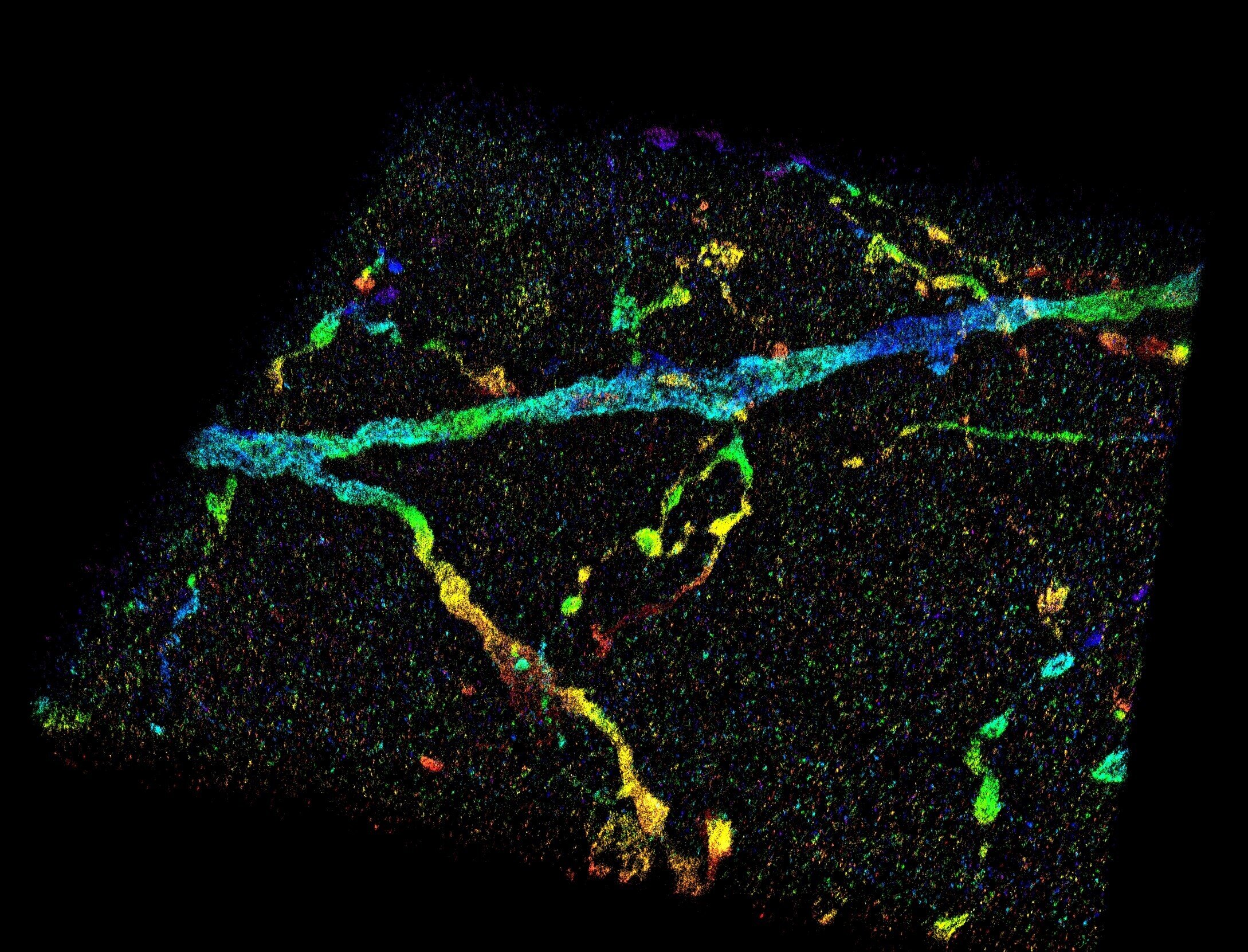
A super resolution 3D reconstruction of dendrites in the primary visual cortex. Innovators at Purdue University created an imaging tool that enables the visualization of nanoscale structures within cells and entire tissues. Credit: Fang Huang / Purdue University
Since Robert Hooke’s first description of a cell in Micrography 350 years ago, microscopy has played an important role in understanding the rules of life.
However, the smallest resolvable characteristic, resolution, is restricted by the wave nature of light. This centuries-old barrier has restricted the understanding of cellular functions, interactions, and dynamics, particularly on a submicron-to-nanometer scale.
Super-resolution fluorescence microscopy exceeds this fundamental limit, offering up to a ten-fold improvement in resolution, and allows scientists to visualize the inner workings of cells and biomolecules at unprecedented spatial resolution.
However, this ability to resolve is impeded when looking at tissue or whole cell samples, such as those often analyzed during brain or cancer studies. Light signals, emitted by molecules within a sample, travel through different parts of cellular or tissue structures at different speeds and cause aberrations, which will deteriorate the image.
Now, researchers at Purdue University have developed new technology to overcome this challenge.
“Our technology allows us to measure sample-induced wavefront distortions, whether it is a cell or a tissue, directly from the signals generated by individual molecules, small light sources attached to the cellular structures of interest,” said Fang Huang , assistant professor of biomedical engineering at the Purdue College of Engineering. “By knowing the induced distortion, we can determine the positions of individual molecules with high precision and accuracy. We obtain thousands or millions of coordinates of individual molecules within a cell or tissue volume and use these coordinates to reveal the nanoscale architectures of the components of the samples. ” “
The Purdue team technology was recently published in Nature’s Methods.
“During super-resolution three-dimensional imaging, we recorded thousands to millions of emission patterns of individual fluorescent molecules,” said Fan Xu, a postdoctoral associate in Huang’s lab and co-author of the publication. “These emission patterns can be thought of as random observations at various axial positions sampled from the underlying three-dimensional point scattering function that describes the shapes of these emission patterns at different depths, which we intend to recover. Our technology uses two steps: mapping and updating , to iteratively retrieve wavefront distortion and three-dimensional responses from the single-molecule dataset containing emission patterns of molecules at arbitrary locations. “
Purdue technology enables biomolecule positions to be found with a precision of a few nanometers within cells and entire tissues, and therefore solve tissue and cell architectures with high resolution and fidelity.
“This advancement extends the routine applicability of super resolution microscopy from selected cell targets near coverslips to intra and extracellular targets within tissues,” said Donghan Ma, a postdoctoral researcher in Huang’s lab and co-author of the publication. . . “This new visualization ability could allow a better understanding of neurodegenerative diseases like Alzheimer’s and many other diseases that affect the brain and various parts of the body.”
The National Institutes of Health provided significant support for the research.
Other members of the research team include Gary Landreth, professor at Indiana University School of Medicine; Sarah Calve, associate professor of biomedical engineering at Purdue College of Engineering (currently associate professor of mechanical engineering at the University of Colorado Boulder); Peng Yin, professor at Harvard Medical School; and Alexander Chubykin, assistant professor of biological sciences at Purdue. The full list of authors can be found at Nature’s Methods.
“This technical advance is surprising and will fundamentally change the precision with which we assess the pathologic features of Alzheimer’s disease,” said Landreth. “We can see smaller and smaller objects and their interactions with each other, which helps reveal structural complexities that we haven’t appreciated before.”
Calve said the technology is a step forward in regenerative therapies to help promote repair within the body.
“This development is critical to understanding tissue biology and being able to visualize structural changes,” said Calve.
Chubykin, whose lab focuses on autism and diseases that affect the brain, said high-resolution imaging technology provides a new method for understanding deficiencies in the brain.
“This is a breakthrough in terms of functional and structural analysis,” said Chubykin. “We can see a much more detailed view of the brain and even mark specific neurons with genetic tools for further study.”
The team worked with the Purdue Research Foundation’s Office of Technology Commercialization to patent the technology. The office recently moved to the Convergence Center for Innovation and Collaboration in the Discovery Park District, adjacent to the Purdue campus.
New development in 3-D super resolution imaging gives insight into Alzheimer’s disease
Three-dimensional nanoscopy of cells and whole tissues with recovery of the point dispersion function in situ, Nature’s Methods (2020). DOI: 10.1038 / s41592-020-0816-x, www.nature.com/articles/s41592-020-0816-x
Provided by
Purdue university
Citation:
Imaging technology enables visualization of nanoscale structures within entire cells (2020, May 4)
Retrieved on May 4, 2020
from https://phys.org/news/2020-05-imaging-technology-visualization-nanoscale-cells.html
This document is subject to copyright. Apart from any fair treatment for the purpose of study or private investigation, no
part may be reproduced without written permission. The content is provided for informational purposes only.
[ad_2]
