. Credit: Rockefeller University")
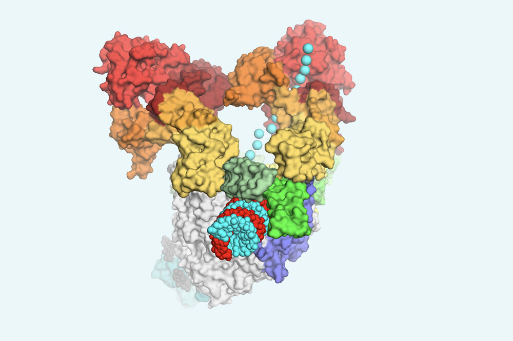
Two essential enzymes coordinate to read and copy SARS-CoV-2 genetic material (displayed as blue spheres). Credit: Rockefeller University
Just how viruses replicate is a complex puzzle with many missing pieces. And in the time of the pandemic, resolving it has become a matter of acute urgency.
Finding out the details of the fundamental copy operations that make one virus in thousands more can give scientists an important leg up in discovering new drugs that contaminate SARS-CoV-2 and stop infection in its tracks.
In a new study published in Sel, Rockefeller scientists provide a crucial piece of the puzzle: an atomic level view of the SARS-CoV-2 replication system. “We now have an additional structural template that can be really useful for drug developers trying to find new compounds that could get into this molecular machine and stop it,” says Elizabeth Campbell, a research assistant at Rockefeller.
Multipart machines
Like many other viruses, the coronavirus copies its genetic material using an enzyme as complex as its name would suggest: the RNA-dependent RNA polymerase, or RdRp. Because it is absolutely essential for viral replication, this machine is thought to be a promising target for antiviral drugs. In fact, some existing antivirals exist, such as several new candidates being tested specifically for COVID-19 on RdRp – including inhibitor, which is currently being used in various countries for the treatment of severe cases.
These antiviral drugs try to hang in necks and crowns of the giant RdRp molecule, like clogs in its gears, causing the machine to stop. To remove this, a connection must be exceptionally precise – which means that scientists trying to design a successful connection need the most detailed picture of the RdRp they can possibly get.
Further complicating matters is the fact that RdRp does not work alone. It does involve a number of other proteins, including another crucial enzyme called helicase, which in its own right is a promising target for COVID-19 drug discovery. This tight cluster of RdRp and associated proteins is “probably what the enzyme looks like outside the lab and in its natural environment, in an infected cell,” says James Chen, a postdoctoral associate in Seth Darst’s lab and one of the first authors of ‘ the study.
Using a powerful imaging technique called cryo-electron microscopy, the team of Darst and Campbell, and their collaborators in the labs of Brian Chait and Tarun Kapoor, were able to show exactly what this multipart machine looks like. One piece of good news: Even if they form a complex, the cavities of the RdRp or the helicase do not change shape, so molecules designed to inhibit these enzymes in isolation may still work on the duo. What’s more, the image reveals several previously unknown sites in the machine that are vulnerable to drugs – including one spot at the interface between the two enzymes, a joint that could potentially be dismantled by an interfering molecule.
The coronavirus crack and more
The new findings could improve human health in several ways. In the case of COVID-19, as scientists around the world race to find antiviral molecules, the new data could significantly accelerate their work. In particular, the unusual resolution at which the team produced its 3-D map of the RdRp helicase complex will support computer studies in which researchers examine the function of drug candidates “purely” based on their knowledge of the chemical structure of the molecules.
“If one is looking for molecules that can stick in a particular binding pocket, having a detailed picture of what that pocket looks like greatly improves the precision of computer docking,” says Brandon Malone, a graduate student at Rockefeller and the co-author of the study.
Beyond COVID-19, the new findings would also help scientists narrow down their ideas on how exactly the two enzymes read and copy genetic material in all so-called RNA viruses, a large group of pathogens including everything from coronaviruses to dengue, Ebola, and the common flu .
“Now we will not only be able to propose models for the mechanics of viral replication, but also actually test these models.” since Chen.
Amid the rush for COVID-19 drugs, a case for the helicase
James Chen et al. Structural basis for helicase polymerase coupling in the SARS-CoV-2 replication-transcription complex, Sel (2020). DOI: 10.1016 / j.cell.2020.07.033
Sel
Delivered by Rockefeller University
Citation: An unprecedented image of the coronavirus copier (2020, August 10) retrieved August 10, 2020 from https://phys.org/news/2020-08-never-before-seen-image-coronavirus-machine. html
This document is subject to copyright. Except for any fair treatment for the purpose of private study or research, no part may be reproduced without the written permission. The content is provided for informational purposes only.
